

TL;DR:
- If you experience a sudden "pop" at the back of your ankle or cannot push off your foot, treat this as a likely Achilles rupture until assessed by a clinician. Clinical signs like an inability to heel raise, palpable tendon gap, and positive Thompson test suggest rupture, whereas gradual pain may indicate tendinopathy. Emergency care is urgent if red flags such as visible gap, severe swelling, or loss of circulation are present.
If you heard or felt a sudden "pop" at the back of your ankle, cannot push off through your foot, or have visible swelling and bruising above your heel, treat this as a probable Achilles tendon rupture until a clinician says otherwise. The Thompson test (calf-squeeze test), Matles test, and Royal London Hospital test are the three clinical checks that matter most, and over 20% of ruptures are initially misdiagnosed because patients can still walk after a complete tear.
Red flags requiring immediate A&E attendance or a 999 call:
- Audible "pop" or sensation of being kicked, with sudden loss of push-off power
- Complete inability to bear weight or stand on tiptoe on the affected leg
- Visible gap or depression in the tendon, roughly 2–6 cm above the heel
- Severe, rapid swelling and bruising spreading around the ankle
- Open wound over the tendon area
- Numbness, tingling, or loss of circulation in the foot
If any red flag applies: immobilise the foot in slight plantarflexion (toes pointing slightly downward), avoid all weightbearing, and go to A&E or call 999. If symptoms are significant but no red flags are present, contact your GP for a same-day appointment or book an urgent physiotherapy assessment at Parkstherapycentre.
Table of Contents
- How to do a step-by-step Achilles injury assessment at home
- What a clinician will do: the full assessment sequence
- When is imaging actually needed?
- How to tell rupture from tendinopathy, bursitis, or an ankle sprain
- What to do immediately after suspecting a serious Achilles injury
- Treatment options and what rehabilitation looks like
- How to prepare for your GP, physio, or orthopaedic appointment
- The Parkstherapycentre clinical perspective on Achilles assessment
- Key takeaways
- Parkstherapycentre: expert Achilles assessment when you need it
- Useful sources and further reading
How to do a step-by-step Achilles injury assessment at home
Safe self-checks cannot replace a clinical diagnosis, but they can tell you quickly whether your symptoms are consistent with a serious rupture or a less urgent tendon problem. Perform these checks on a firm surface, ideally with a partner, and stop immediately if any step causes severe pain or instability.
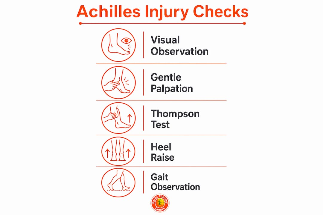
Step 1: Visual observation
Sit on a chair or the edge of a bed and look at both ankles side by side. Note any swelling, bruising, or skin discolouration above the heel. A visible dip or hollow in the tendon line, roughly 4–6 cm above where the tendon meets the heel bone, is a strong indicator of a significant tear. Compare the contour of both Achilles tendons; even slight asymmetry is worth noting.
Step 2: Gentle palpation
With clean hands, run two fingers slowly along the back of the affected ankle, starting at the heel and moving upward. The tendon should feel firm and continuous. A soft, boggy area or a distinct step-change in the tendon's profile, particularly at the 2–6 cm zone above the calcaneal insertion, suggests a gap consistent with rupture. Do not press hard; light pressure is enough to detect a defect.
Step 3: The Thompson (calf-squeeze) test at home
This is the most reliable self-check for a complete rupture.
- Lie face-down on a bed or sofa with your feet hanging off the edge.
- Ask a partner to squeeze the widest part of your calf firmly with both hands.
- Watch your foot: in a healthy tendon, the foot should plantarflex (point downward) in response to the squeeze.
- If the foot does not move, or moves significantly less than the uninjured side, this is a positive Thompson test and strongly suggests a complete rupture.
Do not attempt this test if there is an open wound, severe pain on touch, or any neurovascular concern. The Thompson test carries pooled sensitivity figures of 96–100% and specificity of 93–100% in clinical reviews, making it the most informative single check available outside a clinic.
Step 4: Single-leg heel raise
Stand near a wall for support and try to rise onto the toes of the affected foot alone. A person with an intact Achilles tendon can complete this movement, though it may be painful with tendinopathy. Complete inability to rise, or a very weak, collapsing attempt, is abnormal and warrants urgent assessment. If you can perform the raise but feel pain or stiffness, a partial tear or tendinopathy is more likely than a complete rupture.

Step 5: Gait and weightbearing observation
Walk a few steps if it is safe to do so. A pronounced limp, a flat-footed gait (no push-off), or complete inability to bear weight all increase suspicion of a serious injury. Pain that eases slightly with a short warm-up but returns worse the following day is a classic pattern for Achilles tendinopathy rather than rupture.
Limitations: partial tears and severe tendinopathy can produce false-negative results on all of these checks. A patient with a partial tear may still plantarflex on the Thompson test and may manage a weak heel raise. A history of a "pop" or "kick" sensation should be treated as a rupture until excluded professionally, regardless of what the self-checks show.
Pro Tip: Always compare the injured side with the uninjured side for every check. Asymmetry, not just pain, is the most reliable signal at home.
What a clinician will do: the full assessment sequence
A structured clinical assessment follows a clear sequence. Clinicians form a working hypothesis from the history before they touch the patient, then use hypothesis-driven examination to select the most informative tests.
Subjective history
The clinician will ask about the mechanism of injury (sudden "pop" or gradual onset), whether the sensation felt like a kick from behind, the timeline, prior tendon symptoms, steroid injections near the tendon, use of fluoroquinolone antibiotics (which increase rupture risk), activity level, and relevant comorbidities such as diabetes or inflammatory arthritis. This history guides which physical tests are prioritised.
Lower-quarter screen and observation
Before any specific test, the clinician observes gait, standing posture, foot alignment, and footwear wear patterns. A structured subjective and objective examination that includes biopsychosocial factors is recommended in clinical guidelines. Footwear with excessive heel wear on one side, or a pronated foot posture, can contribute to tendon loading patterns and informs the differential diagnosis.
Key clinical tests: how they are performed and what they mean
Thompson (calf-squeeze) test The patient lies prone with feet off the table. The clinician squeezes the calf. Absence of plantarflexion is a positive result, indicating complete rupture. This is the single most diagnostically powerful test.
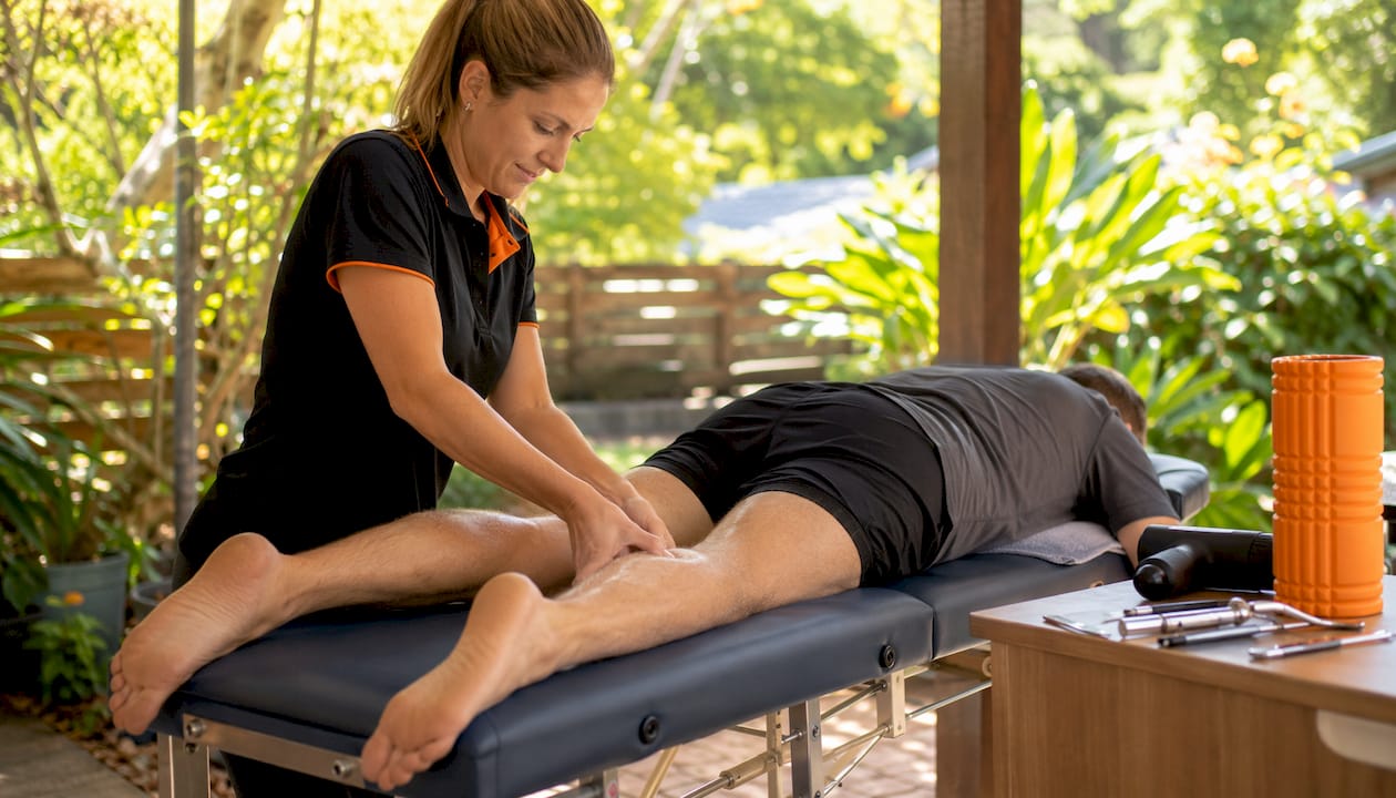
Matles test The patient lies prone and actively flexes both knees to 90°. The clinician observes the resting position of both feet. In a normal tendon, the foot rests in slight plantarflexion. If the affected foot falls into neutral or dorsiflexion relative to the other side, the test is positive for rupture. This test is particularly useful when the Thompson result is equivocal.
Royal London Hospital test The clinician palpates the point of maximum tenderness in the tendon with the ankle in plantarflexion. The foot is then moved into dorsiflexion. If the pain reduces or disappears as the tendon is stretched over the palpation point, the test is positive for mid-portion tendinopathy. This distinguishes tendinopathy from other causes of posterior ankle pain.
Arc sign The clinician asks the patient to actively move the ankle through plantarflexion and dorsiflexion while palpating a tender nodule on the tendon. If the nodule moves with the tendon (arcs with movement), this indicates the pathology is within the tendon body itself, consistent with tendinopathy. A fixed nodule suggests paratendinopathy or another structure.
Palpation for tendon gap Direct palpation along the tendon identifies a step-off or gap, most commonly 2–6 cm above the calcaneal insertion. Palpation for tendon thickening and crepitus is also a strong diagnostic sign for mid-portion tendinopathy.
Single-leg heel raise and hop test These tendon-loading measures have high specificity as diagnostic tests, meaning a positive result (failure to complete) strongly supports the diagnosis. However, their sensitivity is lower, so a patient who manages a weak raise cannot be assumed to have an intact tendon. The heel-raise test assesses calf endurance and motor control rather than maximal strength alone.
Diagnostic accuracy summary
| Clinical test | What it detects | Diagnostic strength |
|---|---|---|
| Thompson (calf-squeeze) | Complete rupture | Very high sensitivity and specificity |
| Matles | Complete rupture | High; useful when Thompson equivocal |
| Royal London Hospital | Mid-portion tendinopathy | Moderate; distinguishes tendinopathy from other pain |
| Arc sign | Tendon body pathology vs paratendinopathy | Moderate specificity |
| Palpation for gap | Complete rupture | High when gap clearly palpable |
| Single-leg heel raise | Tendinopathy / partial tear severity | High specificity, lower sensitivity |
| Hop test | Tendinopathy / partial tear severity | High specificity, lower sensitivity |
Data drawn from pooled systematic review findings; individual study figures vary.
Clinical signs that increase suspicion of complete rupture:
- Positive Thompson test (absent plantarflexion)
- Positive Matles test (foot drops to neutral or dorsiflexion)
- Palpable tendon gap
- Inability to perform a single-leg heel raise
- History of sudden "pop" or "kick" sensation
When is imaging actually needed?
Clinical assessment is often more accurate than imaging for acute Achilles tendon ruptures. In UK practice, MRI and ultrasound are not mandatory for an initial diagnosis when the clinical picture is clear. Imaging is reserved for specific situations.
When imaging adds value:
- Ambiguous clinical findings (equivocal Thompson or Matles test)
- Suspected partial tear where extent of damage affects management decisions
- Subacute or chronic presentations where tendon quality and gap size matter for surgical planning
- Ruling out other pathologies (posterior ankle impingement, os trigonum, peroneal tendon tear)
- Preoperative planning to assess gap size and tendon quality
Ultrasound vs MRI:
Ultrasound is the first-line imaging choice in most UK departments for Achilles pathology. It is quick, relatively inexpensive, and can differentiate partial from complete tears in real time. Ultrasound accuracy is operator-dependent, which is a genuine limitation. MRI provides superior soft-tissue detail, is less operator-dependent, and is preferred when the ultrasound result is inconclusive or when detailed preoperative anatomy is required.
Pro Tip: If a clinician tells you imaging is not needed for an acute, clear-cut rupture, this is evidence-based practice, not a shortcut. A combination of the Thompson, Matles, and palpation for tendon gap has very high sensitivity for complete ruptures, and imaging rarely changes immediate management in those cases.
How to tell rupture from tendinopathy, bursitis, or an ankle sprain
Posterior ankle pain has several causes, and the assessment findings above help distinguish between them.
Quick-reference symptom table:
| Symptom / finding | Most likely diagnosis |
|---|---|
| Sudden "pop," positive Thompson, palpable gap | Complete Achilles rupture |
| Gradual onset, pain 2–6 cm above heel, worse after rest | Mid-portion tendinopathy |
| Pain at heel bone insertion, worse in the morning | Insertional tendinopathy |
| Crepitus, diffuse swelling around tendon, no nodule | Paratendinopathy |
| Swelling at heel, pain with shoe pressure, no arc sign | Retrocalcaneal bursitis |
| Lateral ankle swelling, tenderness over lateral ligaments | Ankle sprain |
Key differentiators at a glance:
- Rupture: sudden onset, "pop" or "kick" sensation, positive Thompson, palpable gap, inability to heel raise.
- Mid-portion tendinopathy: gradual onset, localised nodule 2–6 cm above insertion, positive Royal London Hospital test, positive arc sign, pain that improves with warm-up but worsens the day after loading.
- Insertional tendinopathy: pain at the calcaneal insertion, aggravated by hill running or stiff-soled shoes, no arc sign.
- Paratendinopathy: diffuse, circumferential swelling and crepitus; the arc sign is negative (nodule does not move with tendon).
- Retrocalcaneal bursitis: swelling sits between the tendon and the heel bone; direct pressure from footwear aggravates it; no tendon gap.
- Ankle sprain: tenderness over the lateral or medial ligaments, not the tendon; anterior drawer test positive; Thompson test normal.
For a broader overview of posterior ankle pain causes, the pattern of onset and pain location are the most reliable starting points.
What to do immediately after suspecting a serious Achilles injury
Immediate first aid
- Stop all activity and avoid weightbearing on the affected leg.
- Position the foot in slight plantarflexion (toes pointing gently downward) to reduce tension on the tendon ends.
- Apply a bag of ice wrapped in a cloth for up to 20 minutes to reduce swelling; do not apply ice directly to skin.
- Take paracetamol for pain relief if there are no contraindications; avoid non-steroidal anti-inflammatory drugs (NSAIDs) in the first 48–72 hours if a complete rupture is suspected, as inflammation may play a role in early healing.
- Improvise a splint or use a firm boot if available to maintain the plantarflexed position.
UK care pathways
- Call 999 or go to A&E immediately if there is an open wound, neurovascular compromise, or complete inability to bear any weight with a clear "pop" history.
- Contact your GP for a same-day appointment if red flags are absent but the Thompson test is positive or a gap is palpable; request an urgent orthopaedic or physiotherapy referral.
- Book an urgent physiotherapy assessment at Parkstherapycentre if symptoms suggest a partial tear or tendinopathy without red flags; a physiotherapist can perform the full clinical battery and advise on imaging.
What to bring to your appointment
- A brief written timeline: when the injury happened, what you were doing, and what you felt.
- Photos of swelling or bruising taken in the first 24–48 hours.
- The footwear you were wearing at the time of injury.
- A list of current medications, particularly steroids or fluoroquinolone antibiotics.
Treatment options and what rehabilitation looks like
Non-operative vs operative care
The decision between conservative management and surgical repair depends on patient age, activity level, the size of the tendon gap, and how quickly the injury is assessed. Both approaches are supported by evidence for complete ruptures; the choice is made jointly by the patient and their clinical team.
Non-operative management involves immobilisation in a plantarflexed position, typically in a functional brace or cast, followed by progressive loading under physiotherapy supervision. Re-rupture rates with modern functional rehabilitation protocols are comparable to surgical repair in many patient groups.
Surgical repair (open or percutaneous) reapproximates the tendon ends directly and may be preferred for younger, highly active patients, large gap sizes, or delayed presentations where the tendon ends have retracted. Surgical risks include wound complications and sural nerve injury.
Nutritional support during recovery is worth considering; collagen supplementation and adequate protein intake are increasingly discussed in tendon rehabilitation literature, though individual guidance from your clinician is advisable.
Typical rehabilitation milestones
- 0–2 weeks: Protected immobilisation, swelling management, non-weightbearing or toe-touch weightbearing.
- 2–6 weeks: Gradual progression to full weightbearing in a functional boot; gentle range-of-motion exercises begin.
- 6–12 weeks: Boot weaning, progressive calf strengthening, proprioception work.
- 3–6 months: Return to low-impact activity; single-leg heel raises with increasing load; physiotherapy-guided loading progressions are central to this phase.
- 6–12 months: Return to sport for most patients; full calf strength and power recovery can take up to 12 months or longer.
Common complications to be aware of:
- Re-rupture (higher risk if return to loading is rushed)
- Tendon lengthening (reduces push-off power)
- Persistent calf weakness
- Sural nerve sensitivity (particularly post-surgery)
Achilles injury prevention strategies during and after rehabilitation reduce the risk of recurrence.
How to prepare for your GP, physio, or orthopaedic appointment
What to do before you arrive
- Write down the exact mechanism: "I was sprinting and felt a pop behind my ankle" is far more useful than "my ankle hurts."
- Note the time of injury, any treatments already tried, and whether symptoms have changed.
- Bring the footwear worn at the time of injury; clinicians assess wear patterns as part of the lower-quarter screen.
- List all medications, including any recent courses of corticosteroids or fluoroquinolones.
- Bring photos of bruising or swelling taken in the first 24–48 hours if the appointment is delayed.
What typically happens at the appointment
The clinician will take a brief focused history (roughly 5–10 minutes), then perform a physical examination that includes the tests described above: Thompson, Matles, Royal London Hospital test, arc sign, palpation, and loading tests. They will document findings and, if needed, refer for ultrasound or MRI. You may leave with a diagnosis, a management plan, or a referral letter.
Questions worth asking your clinician:
- Is this likely a partial or complete tear?
- Do I need imaging, and if so, how soon?
- What are the treatment options and their respective timelines?
- When can I bear weight, and what should I avoid in the meantime?
- What are the signs of a complication I should watch for?
The Parkstherapycentre clinical perspective on Achilles assessment
Parkstherapycentre has provided multidisciplinary musculoskeletal care across Bedfordshire and Buckinghamshire since 1986, with physiotherapy and sports injury assessment at the core of its services. The clinic's approach to Achilles assessment follows the structured, hypothesis-driven model described throughout this article: subjective history first, lower-quarter screen, then targeted clinical tests before any imaging decision.
A consistent finding in clinical practice is that patients who can still walk after a rupture are frequently reassured by non-clinical contacts that "it can't be that bad." This is one of the most common pathways to delayed diagnosis. Over 20% of Achilles tendon ruptures are initially misdiagnosed, often precisely because the patient retained some plantarflexion through intact accessory muscles.
"The Thompson test, Matles test, and palpation for a tendon gap together provide a highly sensitive clinical picture for complete rupture. Imaging is valuable in ambiguous cases, but for an acute presentation with a clear history and positive clinical tests, the priority is prompt immobilisation and referral, not waiting for a scan. Treat the 'pop' as a rupture until a clinician tells you otherwise." — Ivan, Parkstherapycentre physiotherapist
Conditions that Parkstherapycentre clinicians frequently see misdiagnosed as Achilles rupture include plantaris tendon injury (a sharp posterior calf pain that can mimic rupture but leaves the Thompson test negative), paratendinopathy, sural nerve irritation, and posterior ankle impingement. A thorough sports injury assessment that includes all four clinical tests, combined with a careful history, reduces the risk of missing any of these.
Key takeaways
A positive Thompson test combined with a palpable tendon gap and a history of a sudden "pop" is the strongest clinical indicator of complete Achilles rupture, and imaging is not required to act on that finding in UK practice.
| Point | Details |
|---|---|
| Treat a "pop" as rupture | A sudden pop or kick sensation should be managed as a rupture until a clinician formally excludes it. |
| Thompson test is the priority check | Absent plantarflexion on calf squeeze is the single most reliable sign of complete rupture. |
| Clinical exam before imaging | UK practice reserves ultrasound and MRI for ambiguous, subacute, or preoperative cases, not routine acute ruptures. |
| Know the red flags | Inability to bear weight, palpable gap, and positive Thompson test warrant same-day urgent assessment or A&E. |
| Parkstherapycentre for formal assessment | Parkstherapycentre offers structured physiotherapy and sports injury assessment across Bedfordshire and Buckinghamshire for patients who need a full clinical evaluation. |
A clinician's note on common assessment errors
The most consequential mistake in Achilles assessment is equating movement with tendon integrity. Partial tears and even some complete ruptures allow residual plantarflexion through the long toe flexors and peroneal muscles, so a patient who can move their foot is not automatically safe to discharge without further testing. The clinical tests described here, particularly the Thompson and Matles combination, exist precisely to bypass that ambiguity. If you suspect a serious Achilles injury, the right response is prompt, structured assessment, not watchful waiting. Parkstherapycentre's physiotherapy team is trained to perform this full assessment sequence and advise on the appropriate next steps, whether that is conservative management, imaging, or an orthopaedic referral.
Parkstherapycentre: expert Achilles assessment when you need it
Recovering from a suspected Achilles injury starts with getting the right diagnosis quickly, and that is exactly where Parkstherapycentre's physiotherapy and sports injury team can help. Rather than waiting weeks for a GP referral or an NHS imaging slot, you can access a structured clinical assessment, including the Thompson, Matles, and Royal London Hospital tests, at one of the clinic's Bedfordshire or Buckinghamshire locations.
The first appointment covers a full subjective history, physical examination, and a clear management plan. Where ultrasound is clinically indicated, the team can advise on the fastest route to imaging and coordinate with orthopaedic services if surgical referral is needed. Parkstherapycentre accepts most major insurance providers and offers private self-pay appointments with no long waiting periods.
Book your assessment online or contact the clinic directly to discuss your symptoms. If red flags are present, go to A&E first; for everything else, early physiotherapy assessment gives you the clearest picture and the fastest route to recovery.
Useful sources and further reading
The following sources informed this article and are recommended for readers who want to review the primary evidence:
- Achilles tendon rupture, StatPearls (NCBI Bookshelf): A comprehensive clinical reference covering diagnosis, misdiagnosis rates, and management; particularly useful for understanding why clinical assessment takes priority over imaging in acute presentations.
- The utility of clinical measures for the diagnosis of Achilles tendon injuries: a systematic review with meta-analysis (PMC): The key systematic review underpinning the diagnostic accuracy data for Thompson, Matles, arc sign, Royal London Hospital test, and loading tests; essential reading for understanding the evidence behind each test.
- Achilles tendon tears, Merck Manual (Professional): Authoritative clinical reference for test procedures, imaging indications, and treatment options.
- Achilles tendinopathy toolkit: clinical evaluation, Physio-pedia: A structured clinical toolkit covering palpation, Royal London Hospital test, arc sign, and biopsychosocial screening; practical for clinicians and informed patients alike.
- Expert insights into Achilles tendinopathy assessment, Physio Network: Clinician-focused discussion of hypothesis-driven examination, pain behaviour patterns, and the distinction between tendinopathy and other posterior ankle pathologies.
This article provides general health information and is not a substitute for professional medical advice. Always confirm your diagnosis and management plan with a qualified clinician or your GP.